Key Takeaways
- Keratoconus is a progressive eye condition in which the cornea gradually thins and bulges into a cone shape, scattering light and causing increasingly distorted vision.
- Common symptoms include blurry vision, glare, halos around lights, and frequent prescription changes; many patients initially mistake these signs for standard astigmatism.
- The exact cause is not fully understood, but genetics, eye rubbing, and weakened corneal collagen are believed to play significant roles.
- Keratoconus is diagnosed through a comprehensive eye exam that includes corneal topography, a detailed map of the cornea's shape.
- Modern treatment options, including FDA-approved corneal cross-linking, can stop or significantly slow progression before vision loss becomes severe.
- Everett & Hurite Eyecare Specialists is among the very few highly specialized practices in the tri-state area offering FDA-approved keratoconus cross-linking treatment. Request an appointment to protect your corneal health before the condition advances.
What Happens to the Cornea in Keratoconus?
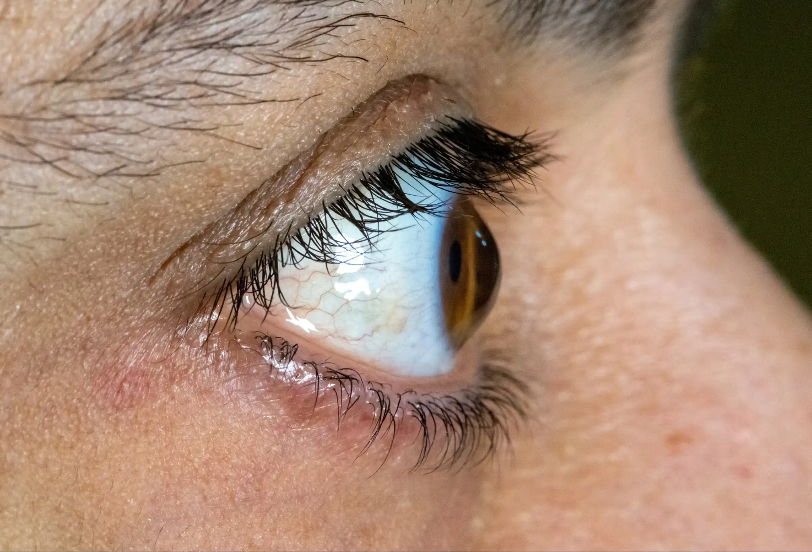
The cornea is the clear, dome-shaped front surface of the eye. Its smooth, rounded shape is what allows light to focus cleanly on the retina at the back of the eye. In keratoconus (pronounced: kair-uh-toe-KOH-nus), the cornea gradually weakens and thins over time. As the tissue loses its structural integrity, it begins to bulge outward into an irregular cone shape instead of holding its normal dome.
That shift in shape means light entering the eye no longer focuses on a single point. Instead, it scatters, causing the kind of blurring, distortion, and halos that glasses alone often cannot fully correct.
According to the American Academy of Ophthalmology, keratoconus affects roughly one in every 2,000 Americans. It most commonly begins in the late teens or early twenties and can progress over the course of 10 to 20 years.
Common Symptoms: Glare, Halos, and Blurry Vision
One of the reasons keratoconus is often missed or misdiagnosed in its early stages is that its initial symptoms overlap significantly with ordinary vision problems like nearsightedness and astigmatism. Warning signs that may indicate keratoconus include:
- Blurry or distorted vision that glasses do not fully correct
- Straight lines that appear wavy or bent
- Glare and halos around lights, especially while driving at night
- Increased sensitivity to bright light
- Frequent and unexplained changes in eyeglass prescription
- Double or ghost images in one eye
- Difficulty seeing clearly at night
- Increasing discomfort with contact lens fit
Because these symptoms tend to develop gradually over years, many patients adapt to subtle vision changes without seeking evaluation. By the time the changes feel obvious, significant corneal changes may already have occurred.
Risk Factors and Possible Causes
The precise cause of keratoconus is still not fully understood. Most evidence points to a combination of genetic susceptibility and environmental triggers that together weaken the cornea's collagen structure over time.
| Risk Factor | What the Research Suggests |
|---|---|
| Family history | First-degree relatives of keratoconus patients have significantly higher risk |
| Eye rubbing | Repeated mechanical stress on the cornea may accelerate thinning |
| Allergies and hay fever | Chronic eye itching and rubbing associated with higher incidence |
| Connective tissue disorders | Conditions like Marfan syndrome and Down syndrome are linked to keratoconus |
| Poorly fitting contact lenses | Chronic pressure may contribute to corneal changes over time |
| Exposure to UV light | Some evidence suggests UV damage may weaken corneal collagen |
A family history of keratoconus is particularly relevant. If a parent or sibling has the condition, a comprehensive corneal evaluation is worthwhile even without noticeable symptoms.
How Keratoconus Is Diagnosed
Standard vision tests measure how clearly you can see, but they cannot detect the irregular corneal shape that characterizes keratoconus. Diagnosis requires a comprehensive eye exam that includes corneal topography, a detailed imaging test that maps the surface curvature of the cornea. Topography allows an ophthalmologist to identify even subtle irregularities that would not appear in a routine refraction.
Additional diagnostic tools may include:
- Corneal pachymetry: Measures corneal thickness at various points to detect thinning.
- Slit-lamp exam: Allows the ophthalmologist to examine the cornea's layers under magnification.
- Wavefront analysis: Evaluates how light travels through the eye's optical system.
Early diagnosis is critical. The earlier keratoconus is identified, the more options a patient has for slowing or stopping its progression before vision loss becomes more difficult to manage.
Modern Treatment Options, Including Corneal Cross-Linking
Treatment for keratoconus depends on the stage of the condition and how quickly it is progressing. In the early stages, glasses or specialty contact lenses may provide adequate vision correction. As the cornea becomes more irregular, standard lenses lose their effectiveness and specialized options become necessary.
Specialty Contact Lenses
For many patients with moderate keratoconus, rigid gas-permeable (RGP) lenses, scleral lenses, or hybrid lenses can vault over the irregular corneal surface and provide significantly clearer vision than soft lenses or glasses. Finding the right lens type requires a specialty contact lens fitting performed by an experienced eye care provider.
FDA-Approved Corneal Cross-Linking
Corneal cross-linking (CXL) is the only treatment shown to stop the progression of keratoconus rather than simply managing its effects. The procedure was approved by the U.S. Food and Drug Administration in 2016 and has since become the standard of care for patients with documented progressive disease.
Cross-linking uses riboflavin (vitamin B2) eye drops combined with ultraviolet-A (UV-A) light exposure to create new bonds between the collagen fibers in the cornea. Those bonds strengthen the corneal tissue, restoring some of its structural integrity and halting further bulging. The procedure typically takes about an hour in-office and is performed under topical anesthesia.
At Everett & Hurite Eyecare Specialists, the practice uses FDA-approved Avedro technology to perform corneal cross-linking. Everett & Hurite is among the very few super-specialized providers in the tri-state area offering this treatment.
Advanced and Late-Stage Options
For patients whose keratoconus has advanced to the point where contact lenses no longer provide adequate vision or are no longer tolerable, surgical options may be considered. These include corneal ring segment implants (Intacs), which help to flatten the cornea's shape, or in the most advanced cases, a corneal transplant. Cross-linking, when performed early enough, can help many patients avoid these more invasive interventions.
Why Early Diagnosis Can Make a Major Difference in Outcomes
Keratoconus is a condition where timing matters enormously. Cross-linking is most effective when performed while the cornea still has adequate thickness and before vision has deteriorated significantly. Patients who are treated early can stabilize their corneas, preserve their existing vision, and continue managing with specialty lenses. Patients who reach advanced stages may find that their options are more limited and that the path to clear vision is considerably more difficult.
The takeaway for Pittsburgh-area patients: if you are experiencing any of the symptoms described above, particularly changing prescriptions, persistent distortion that glasses cannot fix, or halos around lights, a corneal evaluation is warranted. Do not assume the problem is ordinary astigmatism without proper testing.
Get Keratoconus Evaluated at Everett & Hurite
If keratoconus is progressing, early intervention with corneal cross-linking may be the most important step you can take to protect your long-term vision.
Everett & Hurite Eyecare Specialists has nine convenient locations across Pittsburgh, Warrendale, Butler, Greensburg, Southpointe, Monroeville, Steubenville, and Weirton, with a team of ophthalmologists experienced in diagnosing and treating corneal conditions at every stage. Request an appointment and get a comprehensive corneal evaluation today.
Frequently Asked Questions
What is keratoconus?
Keratoconus is a progressive condition in which the cornea, normally a smooth, rounded dome, gradually thins and bulges outward into an irregular cone shape. This changes how light enters the eye and causes increasingly distorted, blurry vision that is difficult to correct with standard glasses.
What are the common symptoms of keratoconus?
Symptoms include blurry or distorted vision, halos and glare around lights (especially at night), increased light sensitivity, frequent prescription changes, and difficulty achieving clear vision with glasses. Many patients also notice that straight lines appear wavy.
Can keratoconus be cured with corneal cross-linking?
Corneal cross-linking does not restore the cornea to its original shape, but it is highly effective at stopping or significantly slowing the condition's progression. For many patients, early treatment with cross-linking allows them to maintain stable vision long-term and avoid more invasive procedures such as a corneal transplant.
What causes keratoconus?
The exact cause is not fully understood. Research points to a combination of genetic factors, weakened corneal collagen, chronic eye rubbing, allergies, and possibly UV light exposure. Having a family member with keratoconus increases risk meaningfully.
Where can I find an eye specialist near me for keratoconus in Pittsburgh?
Everett & Hurite Eyecare Specialists offers FDA-approved keratoconus cross-linking treatment and is one of the few super-specialized providers in the tri-state region for this condition. Nine locations serve patients throughout the Pittsburgh metro area and surrounding states.
